An MRI scan (Magnetic Resonance Imaging) is a highly effective imaging test that helps doctors identify soft tissue damage and other abnormalities. This advanced technology provides detailed images of specific areas, enabling accurate diagnoses for various conditions, including those affecting the ankle joint, leg, and foot.
If you’re experiencing ankle joint pain, foot bone pain, or another discomfort, consider scheduling an MRI scan with the experts at Orthopedic Foot & Ankle Center. Our highly-trained surgeons serve the Greater Columbus area, providing top-tier care for patients with leg and ankle pain.
The very first magnetic resonance imaging exam was performed on a live human on July 3, 1977. Over the past decades, MRI scans have become very useful in helping doctors identify internal areas of concern within the body.
Visiting an orthopedic surgeon doesn’t mean that surgery is necessary. Our orthopedic doctors always prefer to use non-surgical procedures to treat injuries.
“Excellent practice! I would highly recommend them for orthopedic foot and ankle surgery.” — Andrew R.
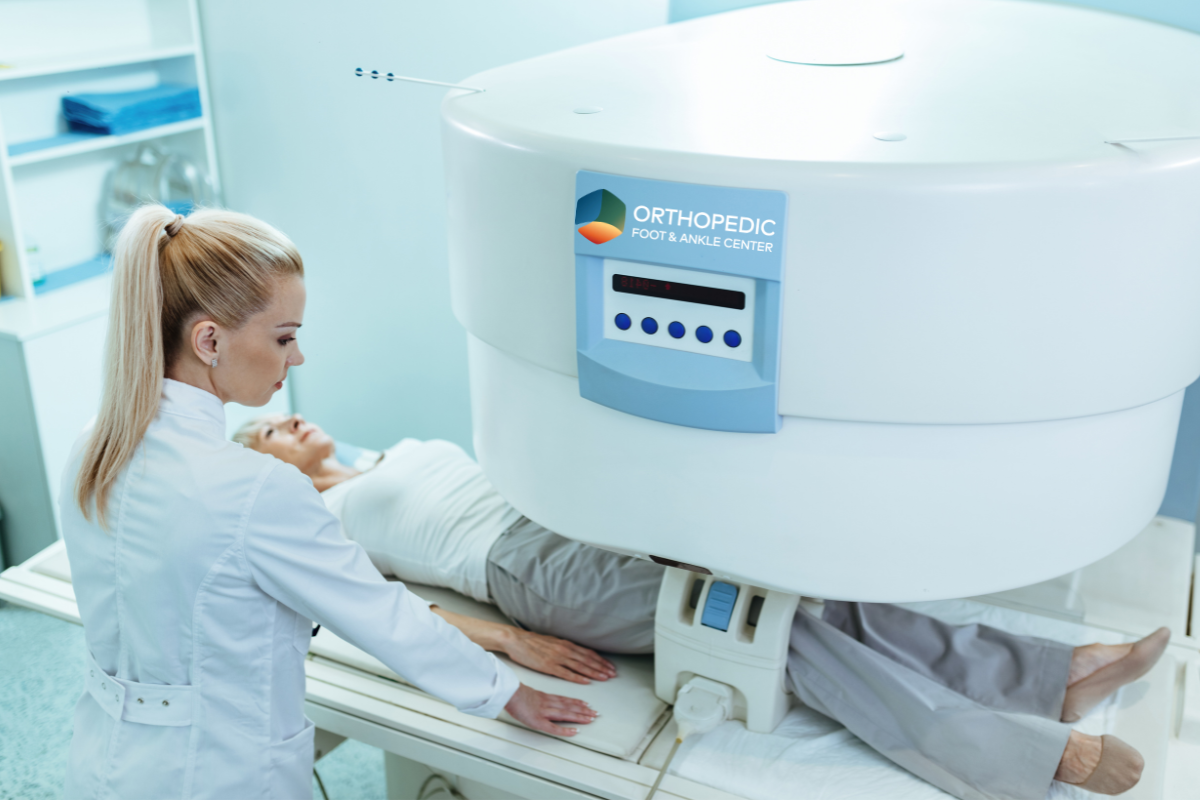
An extremity MRI scan is a specialized imaging test focused on one limb, such as the arm, leg, hand, or foot. The MRI scanner uses radio waves and a magnetic field to generate detailed images of the bones, muscles, joints, blood vessels, and nerves in the area. These images help in diagnosing conditions related to the ankle, foot, and leg.
In comparison to a traditional MRI scanner, an extremity MRI scanner is smaller in size. However, the size does not affect the image quality of extremity MRIs.
The scanner offers a complete MRI system in a compact machine with no RF shielding cage required. They are ideally suited for small diagnostic practices; however, they can also be found in large radiology departments.
An extremity MRI scan is invaluable for diagnosing a range of conditions, especially those related to the foot, leg, and ankle. At Orthopedic Foot & Ankle Center, our surgeons use this technology to diagnose:
Many patients feel anxious or claustrophobic during a traditional MRI exam. With an extremity MRI scanner, patients sit comfortably while only the affected limb is placed inside the machine. This makes the process less stressful and more efficient.
Unlike X-rays, MRI scans do not emit radiation and provide more detailed images of soft tissues, bones, and ligaments, making them ideal for diagnosing problems with the ankle joint, leg pain, foot bone pain, and arthritis.
Additionally, extremity MRI scanners consume less energy than full-body scanners, making them more cost-effective and environmentally friendly.
For most MRI exams, including extremity MRI, no special preparation is necessary. You may be asked to wear loose-fitting clothing or a hospital gown during the procedure. Additionally, remove any metal objects, such as jewelry, implants, or metal plates, as they can blur MRI images.
You should also tell your provider if you have:
The O-scan extremity MRI scanner has an accompanying chair where you can sit comfortably. During the scan, your operator will sit at the console either inside or outside the scanning room.
From your chair, the scanner will be positioned so your limb can be placed inside the scanner. The machine may make a thumping or tapping noise as the magnetic field is turned on and surrounds your limb. You will not feel the magnetic field or radio waves during the scan.
An extremity MRI usually takes about half an hour, though some may last longer. After the procedure is complete, you will be able to return to regular activities.
An extremity MRI provides a safe and comfortable way of providing detailed images of your soft tissues. The resulting images can help your doctor to make an accurate diagnosis of your injury or condition.
While MRI is excellent for diagnosing soft tissue damage, other imaging methods like a bone scan or a leg positron emission tomography (PET scan) may be used for more comprehensive imaging of bone and tissue issues.
If you’re experiencing ankle pain, leg pain, or have concerns about a broken bone, contact Orthopedic Foot & Ankle Center to schedule your leg MRI or foot MRI. Our specialists in Columbus, Ohio, use the latest technology, including extremity MRI, to provide accurate diagnoses for conditions affecting the bones, muscles, and ligaments.
Medically reviewed by Justin R. Hudson, DPM, CWS